|
Macromolecular Crystallography Facility: Home |
|
A more detailed hardware description
Generic background on X-ray tubes (via Wikipedia)
and the origin of the "Kα" notation
for the CuKα X-rays (1.5418 Å) produced by our copper rotating anode targets.
Old System
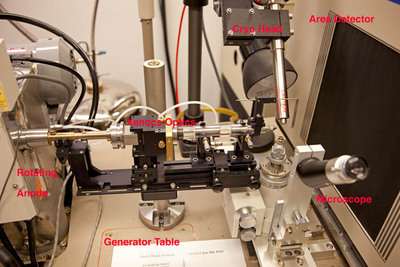 After 17 years we retired a Rigaku RuH3R generator, Xenocs FOX 2D 25-25P
multilayer optic, single axis goniostat, RAXIS IV++ image plate
detector. The RuH3R is what I call a "big spot" rotating anode X-ray generator with an 0.3 x 3mm
focal spot that is much larger than modern microfocus generators - by
about 16-fold larger than the MicroMax 007HF. With the 0.3mm cathode
in place the maximum power was 5.4 kW.
After 17 years we retired a Rigaku RuH3R generator, Xenocs FOX 2D 25-25P
multilayer optic, single axis goniostat, RAXIS IV++ image plate
detector. The RuH3R is what I call a "big spot" rotating anode X-ray generator with an 0.3 x 3mm
focal spot that is much larger than modern microfocus generators - by
about 16-fold larger than the MicroMax 007HF. With the 0.3mm cathode
in place the maximum power was 5.4 kW.
The system was originally supplied with MSC/Yale mirrors (see
"toroidal mirrors" section of Arndt & Bloomer (1999) and in Garman & Sweet (2007)), but a 2012 upgrade
changed the optics from those mirrors to the Xenocs
FOX 2D 25-25P multilayer optic which got us about 3-fold in
intensity gain and rather less CuKβ in our CuKα X-rays.
Nevertheless a larger focal spot inherently begets a larger (or less
brilliant) X-ray beam at the sample.
The Rigaku RAXIS-IV++ detector (see link for Rigaku Journal article on RAXIS-IV, see also Dauter and Wilson (1994)) was a dual image plate
system where the X-ray signal was accumulated in a Eu3+
doped barium halide detector and read out by laser - a lot of moving
parts in this detector but well-engineered and quite reliable until
right at the point where we retired it. Two image plates attached to
a flexible metal belt allowed data collection to continue on one plate
(held flat) while the data was being read out by a laser scanning over
the other plate (held on a cylindrical surface). Moving laser and
laser optics made for a relatively slow readout time (~90 seconds)
which was nevertheless a substantial improvement over previous
versions of IP detectors (8 minutes and 3 minutes) - far slower than a
CCD or CMOS detector. Features included a large active area of 300 x
300 mm, small pixel size (we used 100µ) and high dynamic range.
Nevertheless the detector wasn't very sensitive with a detective quantum efficiency (DQE) of around 20%.
Large area and relatively moderate intrinsic noise led it to dominate
the macromolecular crystallography field for many years and its
production was only recently ceased. Many still use one.
New System
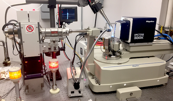 Generator: The MicroMax 007HF is Rigaku's relatively standard
lab rotating anode microfocus source (vendor site) and is the latest generation of the
original microfocus rotating anode generator. Presumably the name
arises from the 0.07 x 0.7 mm small focal spot size of the electron
beam on the rotating anode rather than any affiliation with (e.g.)
Daniel Craig. Viewed from the traditional 6° take-off angle the
rectangular focal spot appears square. The focal spot is 16x smaller
in area than the focal spot for the old RuH3R but runs at 22% of the
power rating (1.2 kW versus 5.4 kW) giving a much brighter focal spot,
and the smaller spot also allows a greater proportion of the X-rays to
be acquired by the X-ray optic (see below) and an inherently smaller
X-ray beam at the sample. Given that most protein crystals are closer
to the 189 µm beam from the generator+optics pairing than they
are to the 300 µm beam from the old RuH3R/Xenocs pair the gain
of the smaller brighter beam is obvious. If you universally had
larger crystals the difference would be much narrower. Rigaku also
make the higher power and larger FR-X rotating anode running at 12,000
rpm and 3 kW if you're up for the additional expense and maintenance
overhead.
Generator: The MicroMax 007HF is Rigaku's relatively standard
lab rotating anode microfocus source (vendor site) and is the latest generation of the
original microfocus rotating anode generator. Presumably the name
arises from the 0.07 x 0.7 mm small focal spot size of the electron
beam on the rotating anode rather than any affiliation with (e.g.)
Daniel Craig. Viewed from the traditional 6° take-off angle the
rectangular focal spot appears square. The focal spot is 16x smaller
in area than the focal spot for the old RuH3R but runs at 22% of the
power rating (1.2 kW versus 5.4 kW) giving a much brighter focal spot,
and the smaller spot also allows a greater proportion of the X-rays to
be acquired by the X-ray optic (see below) and an inherently smaller
X-ray beam at the sample. Given that most protein crystals are closer
to the 189 µm beam from the generator+optics pairing than they
are to the 300 µm beam from the old RuH3R/Xenocs pair the gain
of the smaller brighter beam is obvious. If you universally had
larger crystals the difference would be much narrower. Rigaku also
make the higher power and larger FR-X rotating anode running at 12,000
rpm and 3 kW if you're up for the additional expense and maintenance
overhead.
Optics: We have a Varimax HF optic. Osmic was one of the
earlier companies involved in making multilayer optics: see the "caveat emptor" paper by Yang, Courville and
Ferrara back from the days of Molecular Structure Corporation
before Rigaku acquired both MSC and Osmic - and obviously Rigaku's
VariMax line of multilayer optics are descendents of the original Osmics.
Multilayers are synthetic 2D crystals on an elliptically-curved
substrate where the spacing between the layers in these "crystals"
varies along the length of the optic (See Optics section of Garman and Sweet paper, and
technical discussion via the Rigaku Journal paper by Shimizu & Omote).
Elliptical multilayers with graded spacing are current state of the
art for home source optics with the ability to capture more of the
X-rays out of the source while delivering a parallel or focused X-ray
beam to the source, depending on hardware design. We opted for Rigaku
Varimax HF optics (vendor site) with the Arc)Sec specification
(better polishing) that delivers a beam that is 189µm at FWHM at
the sample. You can buy an optic with a smaller beam at the sample
(VHF model) at the expense of a more divergent beam, or a larger beam
with less divergence (HR model) - yet another example of no free
lunch. The optic is "double bounce" where the X-ray beam bounces off
two orthogonal optical elements. Their action as a 2D diffraction
grating also helps to substantially reduce the CuKβ component to
the desired CuKα X-rays. Other companies also offer graded
multilayer optics of comparable design, including the supplier of our
older multilayer (Xenocs).
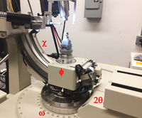 Goniostat: We have a Rigaku AFC-11 partial 4-axis goniostat to
drive the crystal and detector around. Two of the four axes are used
for crystal positioning (φ and χ), one is used for the data
collection scans (ω) and one is used to position the detector
(2θ). There's also a motorized detector distance track. This
sort of goniostat is desired because of the potential for high
resolution, and also to handle the smaller active area of the X-ray
detector (see below) - more crystal permutations are necessary to
achieve desired data completeness. The relatively light detector
allows movement from +5° to -90° in 2θ and the limit on
high resolution data is 0.83 Å due to the CuKα wavelength of
1.54 Å. Your protein crystals will not diffract that far, but
small molecule samples routinely do, especially when mounted on a
powerful rotating anode generator. The φ axis rotates through
360°, ω through a 180° range either side of the 2θ
axis, χ from 0 to 60° and 2θ from +5 to -90°.
Detector to sample distance is in the range 30 to 290 mm. The AFC-11
is relatively slow and not all that exciting from a hardware
perspective and superceded by a faster goniostat after Rigaku's
acquisition of Oxford Diffraction.
Goniostat: We have a Rigaku AFC-11 partial 4-axis goniostat to
drive the crystal and detector around. Two of the four axes are used
for crystal positioning (φ and χ), one is used for the data
collection scans (ω) and one is used to position the detector
(2θ). There's also a motorized detector distance track. This
sort of goniostat is desired because of the potential for high
resolution, and also to handle the smaller active area of the X-ray
detector (see below) - more crystal permutations are necessary to
achieve desired data completeness. The relatively light detector
allows movement from +5° to -90° in 2θ and the limit on
high resolution data is 0.83 Å due to the CuKα wavelength of
1.54 Å. Your protein crystals will not diffract that far, but
small molecule samples routinely do, especially when mounted on a
powerful rotating anode generator. The φ axis rotates through
360°, ω through a 180° range either side of the 2θ
axis, χ from 0 to 60° and 2θ from +5 to -90°.
Detector to sample distance is in the range 30 to 290 mm. The AFC-11
is relatively slow and not all that exciting from a hardware
perspective and superceded by a faster goniostat after Rigaku's
acquisition of Oxford Diffraction.
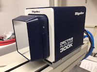 Detector: Dectris Pilatus3 R 300K (vendor site, more technical data). The 300K has three of the
modules, one more than the base 200K detector offered by Rigaku. It's
a photon-counting Hybrid Pixel Array Detector (HPAD) with direct
detection of X-ray photons in the Si detection layer followed by
charge transfer via Indium bump-bonds to an underlying CMOS chip. The
300K uses room-temperature water cooling, and a dry air/nitrogen
supply to suppress humidity. The active area is smaller than the
RAXIS-IV++ (84 mm x 106 mm versus 300 mm x 300 mm) and the pixels are
larger at 172 µm but there are a host of upsides to this
detector: minimal electronic noise; fast readout time (7 millisec);
high dynamic range; single pixel point spread function; very high DQE
(~98% at CuKα); light weight. With the exception of the
relatively small size and the obvious boundaries between the modules
the detector is exceptional and probably accounts for the majority of
the improvement over the old system - perhaps as much as 10x over the
17 year-old RAXIS IV++ we had. Being relatively small and light it
also enables us to collect very high resolution data on e.g. small
molecule samples to around 0.83 Å resolution. CCD and CMOS detectors
of older designs had limited dynamic range and noise issues - the Pilatus
is better than any other detector I've encountered on a home source.
Detector: Dectris Pilatus3 R 300K (vendor site, more technical data). The 300K has three of the
modules, one more than the base 200K detector offered by Rigaku. It's
a photon-counting Hybrid Pixel Array Detector (HPAD) with direct
detection of X-ray photons in the Si detection layer followed by
charge transfer via Indium bump-bonds to an underlying CMOS chip. The
300K uses room-temperature water cooling, and a dry air/nitrogen
supply to suppress humidity. The active area is smaller than the
RAXIS-IV++ (84 mm x 106 mm versus 300 mm x 300 mm) and the pixels are
larger at 172 µm but there are a host of upsides to this
detector: minimal electronic noise; fast readout time (7 millisec);
high dynamic range; single pixel point spread function; very high DQE
(~98% at CuKα); light weight. With the exception of the
relatively small size and the obvious boundaries between the modules
the detector is exceptional and probably accounts for the majority of
the improvement over the old system - perhaps as much as 10x over the
17 year-old RAXIS IV++ we had. Being relatively small and light it
also enables us to collect very high resolution data on e.g. small
molecule samples to around 0.83 Å resolution. CCD and CMOS detectors
of older designs had limited dynamic range and noise issues - the Pilatus
is better than any other detector I've encountered on a home source.
Phil Jeffrey, Princeton, last modified August 8th 2017.